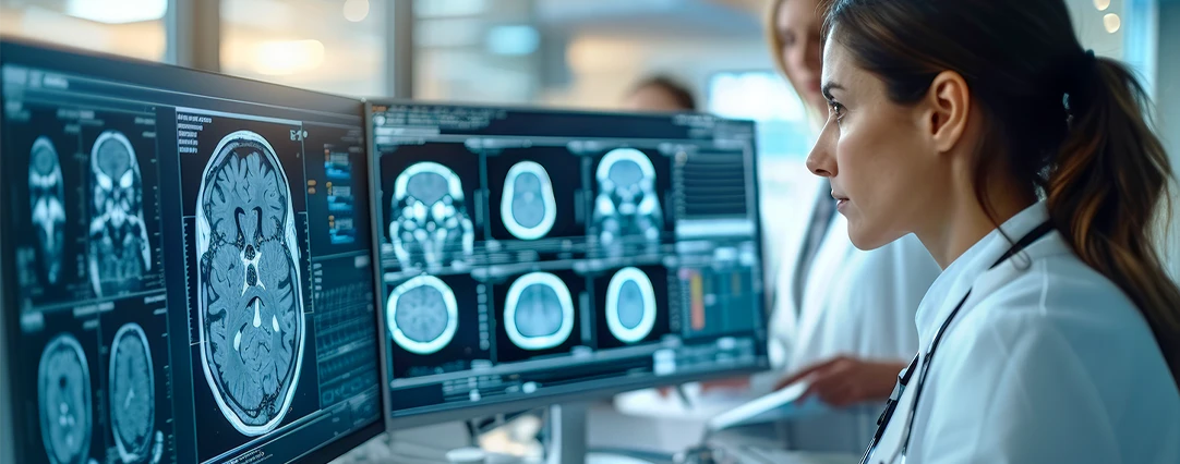
Cardiac imaging specialists use advanced, noninvasive imaging techniques to diagnose and evaluate the treatment of a wide variety of heart and surrounding blood vessel conditions. By using the most advanced, cutting-edge equipment, our cardiac imaging specialists ensure the best diagnostic quality while minimizing the patient’s exposure to radiation.
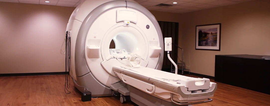
Cardiac imaging uses a variety of imaging equipment, including:
- X-Ray
- CT
- MRI
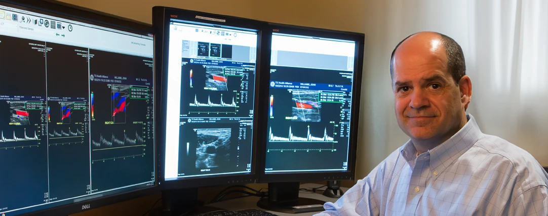
- Echocardiograms
Most cardiac imaging exams use contrast, which is often injected through an arm vein. Contrast helps the radiologist obtain a better view of the heart and blood vessels.

Our Most Common Cardiac Imaging Exam
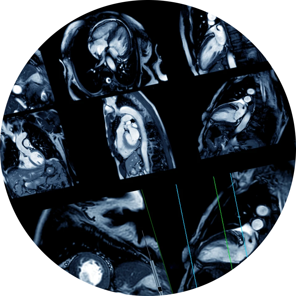
Coronary CT Angiography, also known as Coronary CTA or CCTA, is a noninvasive exam that provides detailed pictures of the heart and its surrounding blood vessels. Radiologists use these images to diagnose conditions, including coronary atherosclerotic disease. The exam can also be performed on advanced MRI machines, which are capable of providing the highest level of detail and specificity in the images.
Exams read by our cardiac imaging specialists
- Echocardiography – uses ultrasound waves to produce an image of the heart and great vessels.
- Chest X-ray – helps doctors view arterial and ventricular size and shape.
- Spiral (or helical) CT – can help evaluate pericarditis, congenital disorders, disorders of the great vessels, tumors, or acute pulmonary embolism.
- Cardiac MRI – This exam does not use X–rays or radiation and can help doctors see the structure and function of the heart muscle.
Cardiac Imaging Section Chief:
Joshua Huff, M.D.

Medical Degree: 2001, University of Texas Southwestern Medical School at Dallas, Texas
Residency: 2006, Diagnostic Radiology, University of Texas Southwestern Medical Center at Dallas, Texas
Fellowship: 2007, MRI University of Wisconsin, Madison, Wisconsin
Board Certified: American Board of Radiology
Meet Our Cardiac Imaging Radiologists

Joshua Huff, M.D.
Medical Degree: Medical Doctor, University of Texas Southwestern Medical School at Dallas, TX (2001)
Residency: Diagnostic Radiology, University of Texas Southwestern Medical Center at Dallas, TX (2002-2006)
Fellowship: MRI University of Wisconsin, Madison, WI (2007)
Board Certified: American Board of Radiology
